Anatomy Of Upper Thigh And Hip - Diagram / Pictures: Neurovasculature of the hip and the ... / Atlas of human anatomy in cross section.
byAdmin•
0
Anatomy Of Upper Thigh And Hip - Diagram / Pictures: Neurovasculature of the hip and the ... / Atlas of human anatomy in cross section.. Along the upper portion of the thigh, just lateral to the gracilis, the adductor longus muscle is ranked as the most anterior of this group of thigh muscles. Medial condyle of tibia nerve supply: Sartorius muscle anatomy page has origin, insertion, innervation, and blood supply information. The hip muscles are going to be slip into hip muscles and gluteal muscles. This deep muscle begins in the low back and pelvis and connects on the inside edge of the upper femur.
The upper part of the aponeurosis is curved backward over the upper edge of the tendon of the gracilis so as to be inserted behind it. Rectus femoris forms the middle portion of the quadriceps. Hip movements include flexion, extension, abduction, adduction, circumduction, and hip rotation. Related online courses on physioplus. Hip anatomy, hip joint, groin anatomy.
Muscles of the Thigh and Gluteal Region - Part 1 - Anatomy ... from i.ytimg.com Unlike the shoulder girdle, the pelvic girdle is firmly integrated into the axial skeleton: Superficial fascia.—the superficial fascia forms a continuous layer over the whole of the thigh; The hip region is located lateral and anterior to the gluteal region, inferior to the iliac crest. Hip flexor deep in pelvis a composite o… used to extend the hip when climbing st… Mri of upper leg (femur). 340 anatomical structures of the hip region were labeled, accessible on anatomical parts: He also serves the communities of charleston, sc and augusta, ga. The thigh is the area between the hip and the knee joint.
Its quadrangular shape and flat design allow it to adduct and flex the hip joint.
This webpage presents the anatomical structures found on thigh mri. The femur or thigh bone is one of the longest bones in the human body. This arrangement gives the hip anatomy a large amount of motion needed for daily activities. Related online courses on physioplus. It is part of the lower limb. Mri of upper leg (femur). Medial condyle of tibia nerve supply: All of the anatomical parts of the hip work together to enable various movements. Here are a few fundamental moves to try. Knee assessment and hip mechanics online course: Bends (flexion) the thigh at the hip. The thigh is the area between the hip and the knee joint. Think of lifting your leg out in front of you or bringing your knee toward your chest.
Quadriceps, a group of four. Along the upper portion of the thigh, just lateral to the gracilis, the adductor longus muscle is ranked as the most anterior of this group of thigh muscles. Bones of the lower limb. Hip flexor deep in pelvis a composite o… used to extend the hip when climbing st… Pelvis, perineum, hip, and upper thigh.
Athletic Training: Thigh, Hip, Groin, and Pelvis Injuries from 2.bp.blogspot.com In order to help understand the conditions causing hip pain and their surgical treatment, it is important to first have a basic understanding of the anatomy of the hip and how it functions. The hip muscles are going to be slip into hip muscles and gluteal muscles. In vertebrate anatomy, hip (or coxa in medical terminology) refers to either an anatomical region or a joint. Here are a few fundamental moves to try. Hip and knee pain and hip and shoulder pain are. Superficial fascia.—the superficial fascia forms a continuous layer over the whole of the thigh; Pelvis, perineum, hip, and upper thigh. Unlike the shoulder girdle, the pelvic girdle is firmly integrated into the axial skeleton:
Hip movements include flexion, extension, abduction, adduction, circumduction, and hip rotation.
Medial condyle of tibia nerve supply: Hip and knee pain and hip and shoulder pain are. Want to learn more about it? Anterior muscles extend your legs and flex your thighs. Hip flexor deep in pelvis a composite o… used to extend the hip when climbing st… The upper part of the aponeurosis is curved backward over the upper edge of the tendon of the gracilis so as to be inserted behind it. The hip's unique anatomy enables it to be both extremely strong and amazingly flexible, so it can bear weight and allow for a wide range of movement. Exercises that involve knee flexion and hip extension are commonly used to build the hamstring muscles. While the thigh muscles will be slip into the anterior, medial and posterior groups. All of the anatomical parts of the hip work together to enable various movements. Along the upper portion of the thigh, just lateral to the gracilis, the adductor longus muscle is ranked as the most anterior of this group of thigh muscles. The hip muscles are going to be slip into hip muscles and gluteal muscles. In vertebrate anatomy, hip (or coxa in medical terminology) refers to either an anatomical region or a joint.
During hip replacement surgery, your surgeon removes the upper part of your thigh bone, including the femoral head (ball of the hip joint) and a part the upper part of the thigh bone is then exposed, and a series of tools called broaches are introduced one at a time to prepare your thigh bone for a metal. It originates at the anterior inferior iliac spine and just above the acetabulum of the hip bone. Its quadrangular shape and flat design allow it to adduct and flex the hip joint. Pelvis, perineum, hip, and upper thigh. for detailed anatomy of pelvic bones, read anatomy of hip bone.
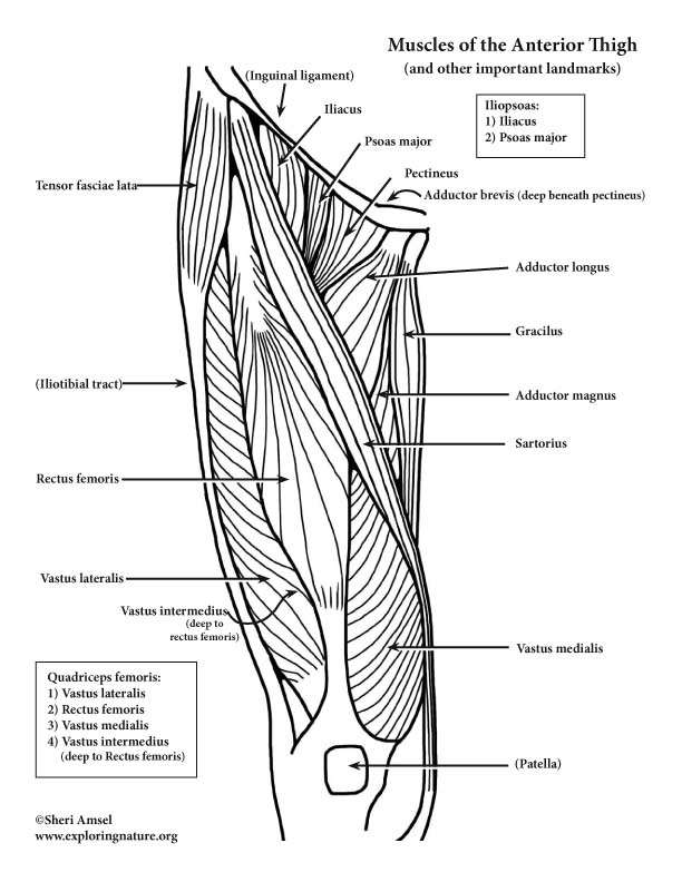
30 Label The Muscles Of The Anterior Thigh. - Labels ... from www.exploringnature.org Its quadrangular shape and flat design allow it to adduct and flex the hip joint. Chief flexor of knee weak. Groin, inguinal region and the anterior and posterior regions of the hip and thigh. Anterior muscles extend your legs and flex your thighs. Hip and knee pain and hip and shoulder pain are. B, muscles of the anterior thigh compartment. Muscles of the hip joint. Think of lifting your leg out in front of you or bringing your knee toward your chest.
Upper part of the ischial tuberosity insertion:
Anterior muscles extend your legs and flex your thighs. The hip's unique anatomy enables it to be both extremely strong and amazingly flexible, so it can bear weight and allow for a wide range of movement. Hip surgeon dr guillaume dumont offers hip pain treatments in columbia, sc. Upper part of the ischial tuberosity insertion: Bends (flexion) the thigh at the hip. The single bone in the thigh region is called the origin: B, muscles of the anterior thigh compartment. Tibial part of the sciatic nerve action: In vertebrate anatomy, hip (or coxa in medical terminology) refers to either an anatomical region or a joint. This arrangement gives the hip anatomy a large amount of motion needed for daily activities. Want to learn more about it? Knee assessment and hip mechanics learn how hip and pelvis mechanics can influence the knee powered by physiopedia start course. Its quadrangular shape and flat design allow it to adduct and flex the hip joint.
The upper part of the aponeurosis is curved backward over the upper edge of the tendon of the gracilis so as to be inserted behind it upper thigh anatomy. Knee assessment and hip mechanics learn how hip and pelvis mechanics can influence the knee powered by physiopedia start course.